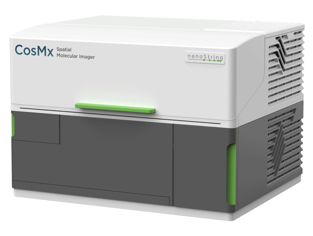
CosMx™ 空間分子イメージャー
CosMx® 空間分子イメージャーのご紹介
空間マルチオミクス シングルセルイメージング プラットフォームのCosMxで、シングルセル研究をステップアップさせましょう
CosMx SMI は、ホルマリン固定パラフィン包埋 (FFPE) および新鮮凍結 (FF) 組織サンプルで、シングルセルおよびサブセルラーの解像度で空間マルチオミクスを提供する、最初のハイプレックス in situ プラットフォームです。 CosMx SMI は、 1,000 の RNA および 100 の検証済みタンパク質分析物の迅速な定量化と可視化を可能にします。 これは、細胞アトラス、組織表現型解析、細胞間相互作用、細胞内プロセス、およびバイオマーカー発見のためのより深い洞察を柔軟に可能とする、シングルセル空間イメージング プラットフォームです。


“We are impressed with ease of use with which the CosMx SMI enabled single cell spatial transcriptomics across various tissue types, even in archival FFPE tissue. The high-plex protein assay makes the CosMx SMI a true multiomic solution. The CosMx SMI’s large capture area allows analysis from valuable clinical trial samples using tissue microarrays, enabling characterization of 100s of samples weekly.”

Dr. Nigel B Jamieson
Professor of Surgery, University of Glasgow, UK
“At last count, we have processed 160 samples with the CosMx SMI, including tumor and mouse brain samples. This technology uncovers vital insights into cell biology, enhancing our understanding of both malignant and normal tissues. It is crucial for creating spatial biomarkers and improving patient outcomes. Plus, the equipment and protocols are reliable and user-friendly.”

Dr. Aubrey Thompson
Mayo Clinic Comprehensive Cancer Center, Florida
“The CosMx SMI complemented our efforts to generate organ atlases of the healthy human system together with our partners of the Human Cell Atlas project. It allowed us to visualize single-cell reference maps directly in organs. The scale of 1000-plex gene panel with image-guided cell segmentation makes the CosMx SMI, a unique platform for spatial genomics!”

Dr. Holger Heyn
Team Leader, Single Cell Genomics Group, CNAG, Spain
“The CosMx SMI instrument has become integral to our laboratory’s future direction, and the responsiveness and skill of the customer support for it and the AtoMx SIP has been absolutely fantastic.”

Dr. Grant Kolar
Director, Research Microscopy and Histology Core, St. Louis University School of Medicine

いち早く体験してください
1000プレックスRNAアッセイによる1回の実験で、包括的な答えを得る

生物学的知見を得るうえで事前設定されたハイプレックスパネルが搭載されています。パネルは、細胞タイプ同定、細胞状態、細胞間相互作用などに利用できる遺伝子を網羅しています。
費用効果の高い構築済みの1000プレックスパネルを適宜カスタマイズして利用することで、あらゆる実験で最大限の結果が得られます。
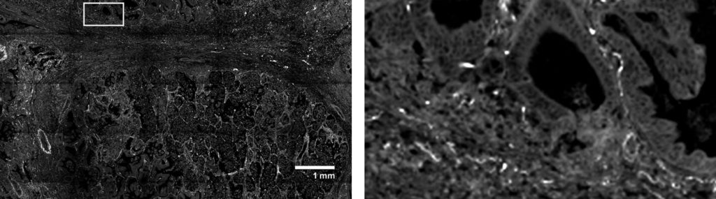
解析が難しいFFPE組織を用いて、空間的状況を考慮しつつシングルセルレベルの遺伝子発現に関する情報を把握する
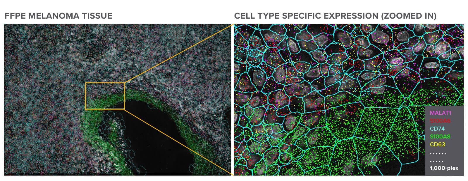
組織検体における転写物の空間的局在を検出するために1000プレックスRNAパネルを用いて探索したFFPEメラノーマ組織
発現量の少ない遺伝子を高感度のCosMx SMIで検出します
| Genes | Expression level | Copies per cell (Max.) |
|---|---|---|
| MALAT1 | High | 256 |
| FOS | Medium | 40 |
| LY6D | Low | 24 |
解析対象とした細胞の総数=800,327個
検出した転写物の総数=259,604,214個
細胞1個当たりの転写物数(最大値)=2,000個
細胞1個当たりの転写物数(平均値)=257個
上に示したのは、FFPE非小細胞肺がん(NSCLC)組織の1000プレックスRNA解析で検出された細胞1個当たりの転写物の数(左)と、NSCLC組織中の転写物の全体的な分布(右)です。CosMx SMIは感度が高く、ダイナミックレンジが広いため、コピー数の少ない遺伝子の転写物でも1細胞レベルで検出できます。
マルチモーダルな手法による本当の意味でのシングルセルセグメンテーション
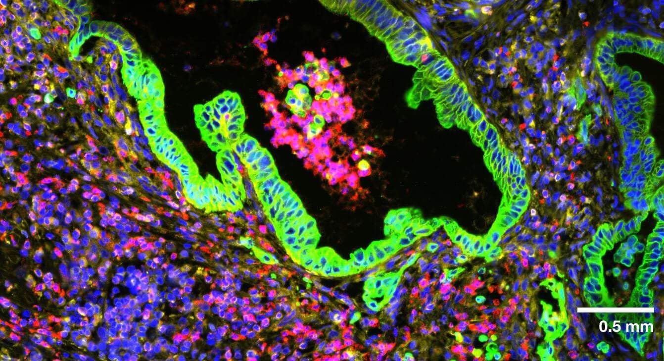

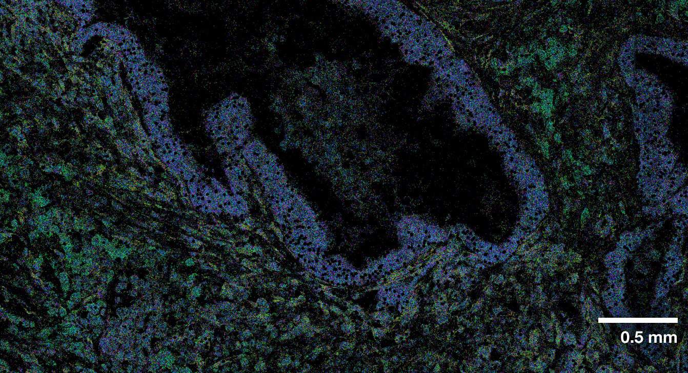
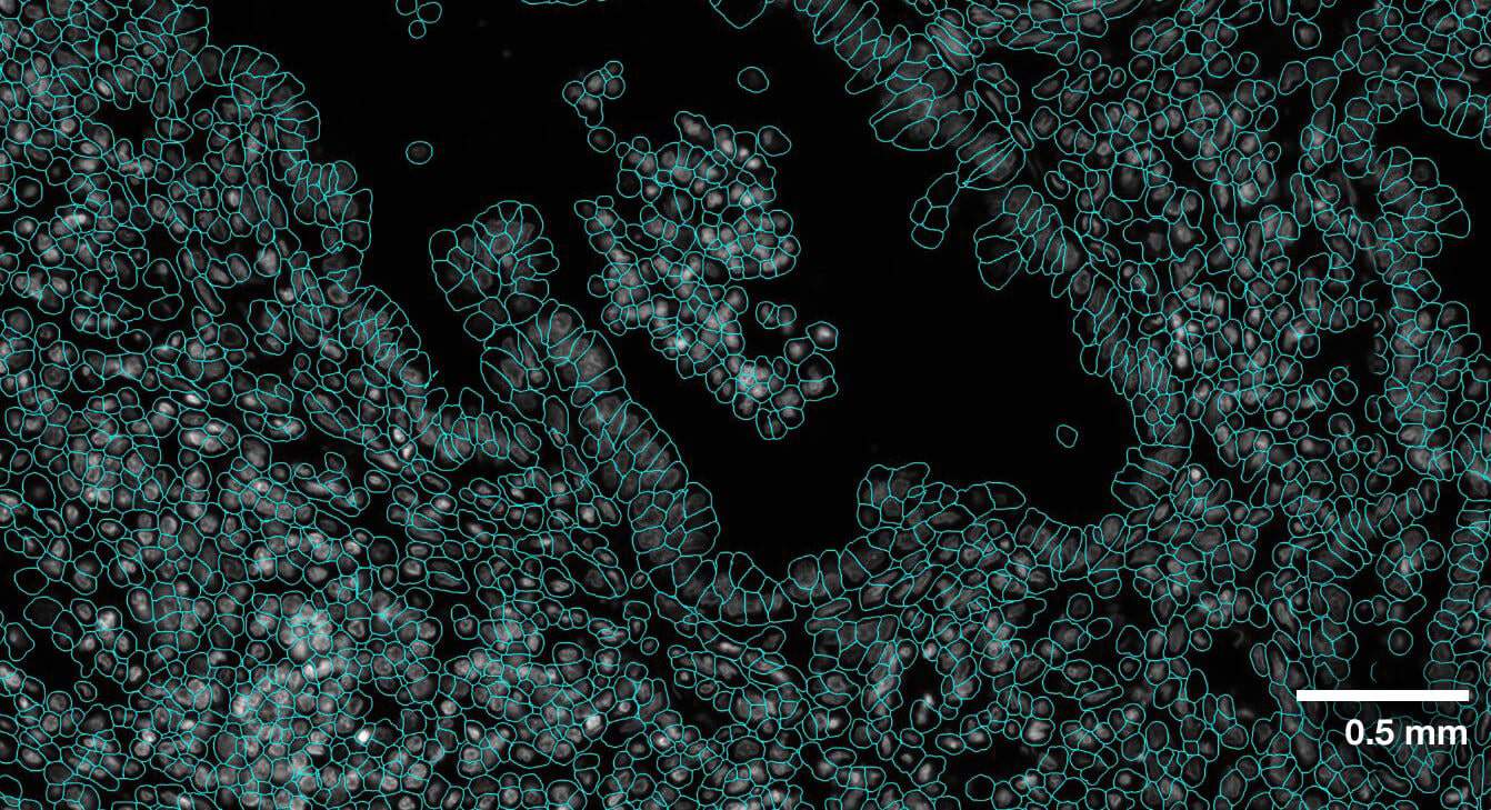
マルチモーダルに細胞領域を分割することで、細胞の境界を正確に検出できます。CosMxの細胞セグメンテーションは、細胞膜と形態マーカータンパク質の画像、機械学習を利用した細胞セグメンテーションアルゴリズム、転写物を基準としたセグメンテーションの精度向上を利用することで、組織の形態を保持したまま正確なシングルセルセグメンテーションを行います
1,000種類ものRNAと100種類ものタンパク質を組織検体で検出可能


RNAだけでなく、100種類に上るタンパク質を組織検体で検出し、空間情報を画像化します。上の画像は、FFPE正常大腸組織において、標的とした7種類のタンパク質を画像化したものです。
CosMx SMI vs. Xenium:
乳癌組織検体での比較解析
本製品でできること
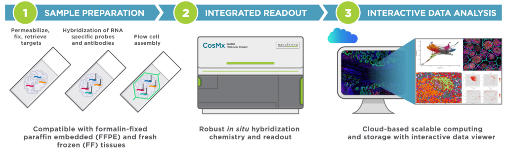
CosMx SMIは、成熟した技術である蛍光in situハイブリダイゼーション(FISH)、高分解能画像のリードアウト、ならびにデータ解析及び画像化のための対話型ソフトウェアを搭載した総合的な解析装置です。
サンプル調製は簡単、どのような種類の検体にも対応
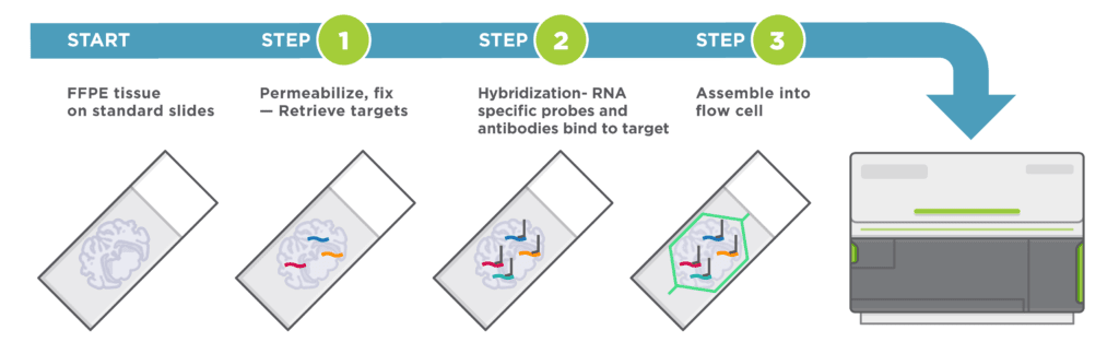
組織の培養や透明化、cDNAの合成や増幅を必要とすることなく、標準的なISHプロトコルに統合可能な、効率化された単純なワークフローです。サンプルから解析結果までの時間を短縮します。
自動サイクルin situハイブリダイゼーション技術
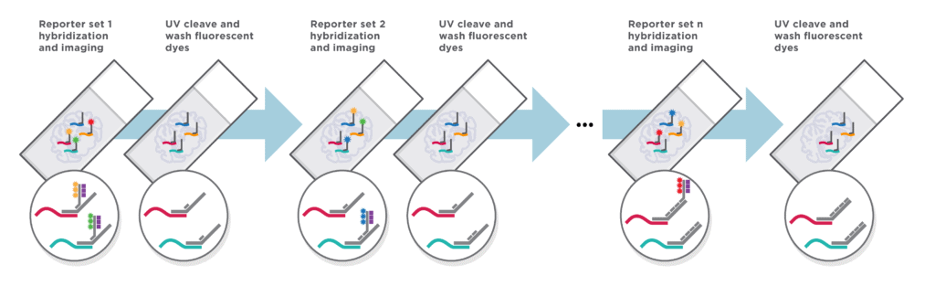
高い感度を実現しハイプレックス解析をサポートする頑健なハイブリダイゼーション技術により、組織検体からこれまで以上の生物学的知見が得られます。
アプリケーション
CosMx空間的分子イメージャーは、以下の目的に使用できる、柔軟性と堅牢性が非常に高い空間生物学のソリューションです。
- 細胞アトラスおよび特性解析:細胞タイプ、細胞状態、組織微小環境の表現型、遺伝子発現ネットワークを空間的に明らかにする。
- 細胞間相互作用:リガンドと受容体の相互作用によって制御される生物学的プロセスを理解する。
- 空間バイオマーカー:空間的状況を考慮しつつ処理による遺伝子発現の変化を定量化し、シングルセルレベルで細胞内バイオマーカーを特定する。


サポート資料
Single-Cell Imaging FAQs
なぜシングルセルが重要なのでしょうか?
細胞は生命の基本的な単位です。 細胞がさまざまな情報層でどのように組織化されて組織を形成するかについての包括的な理解はまだ完全には達成されていません。 さらに、組織がどれほど均一に見えるとしても、そこには多様な細胞集団が含まれており、そのすべてがその組織タイプの異なる症状を表しています。Learn more »
シングルセル解析はどうして重要ですか?
シングルセル解析には、シングルセル解像度でのゲノミクス、トランスクリプトミクス、プロテオミクス、メタボロミクスの研究が含まれます。 細胞は生物の構成要素であるため、さまざまな情報層で組織されて組織を形成し、組織内の各細胞の位置には生理学的または形態学的機能があるからです。 Learn more »
シングルセル技術とは何ですか?
シングルセル技術は単一細胞での操作と増幅における進歩であり、シングルセルのレベルでのゲノミクス、トランスクリプトミクス、およびエピゲノミクスの研究を可能にしました。 Learn more »
シングルセル空間トランスクリプトミクスとは何ですか?
シングルセルレベルでの空間的コンテキストを伴う mRNA 発現プロファイルの解析は、シングルセル空間トランスクリプトミクスとして知られています。 正常状態の細胞と異常状態の細胞の両方において、同様の細胞間でも遺伝子発現パターンが不均一になる可能性があるため、各細胞は固有のトランスクリプトーム フィンガープリントを持っています。 Learn more »
空間トランスクリプトミクスはシングルセルの解像度ですか?
はい。 空間トランスクリプトミクスの分野における最近の進歩により、単一細胞の解像度、場合によっては細胞以下の解像度で RNA 転写物を視覚化することが可能になりました。 Learn more »
シングルセル解析は何に使用されますか?
シングルセル解析では、ゲノム変化、遺伝子発現、環境の影響をシングルセルレベルで研究することで、細胞の表現型に関するデータを得ることができます。 Learn more »