nCounter® Neuropathology Panel
みなさまの研究の一助に
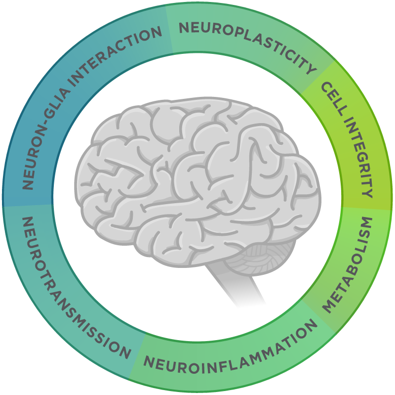
バイオマーカーの発見とシグネチャーの開発は、これまでにない治療法の同定や、アルツハイマー病(AD)、パーキンソン病(PD)、筋萎縮性側索硬化症(ALS)といった神経変性疾患の早期発見や治療法の開発のために欠かせません。nCounter Neuropathology Panelは、神経変性の6つの基本的テーマ(神経伝達、ニューロン・グリア相互作用、神経可塑性、細胞構造の完全性、神経炎症、代謝)に関与する遺伝子を有するヒトまたはマウスのサンプルを対象にした、包括的なマルチプレックス遺伝子発現解析の実施に役立ちます。
- アルツハイマー病、パーキンソン病、筋萎縮性側索硬化症、前頭側頭型認知症、ハンチントン病などの神経疾患の研究向けに開発
- 独自の細胞型特性により、ニューロン、アストロサイト、ミクログリア、オリゴデンドロサイト、内皮細胞などの5つの重要なCNS細胞型の存在量を特定


製品情報
Neuropathology Panelに含まれる遺伝子により、ニューロン、アストロサイト、ミクログリア、オリゴデンドロサイト、内皮細胞などの5つの重要な細胞型の存在量1の測定のための独自の細胞プロファイリングデータを得ることができます。以下の表は、現在の文献で認定されている遺伝子の内容とともに、パネルで提示される各細胞型の概要を示しています。
1 Danaher P. et al. Gene expression markers of Tumor Infiltrating Leukocytes JITC 2017
23の基本的なパスウェイとプロセスの機能アノテーションの割り当ては、Neuropathology Panelの全ゲノムに対して実施されています。これにより、神経変性疾患の発症と進行における重要な側面について実用的な見解を得ることができます。
関連リソース

論文
Spatial transcriptomics reveals molecular dysfunction associated with cortical Lewy pathology
A key hallmark of Parkinson’s disease (PD) is Lewy pathology. Composed of α-synuclein, Lewy pathology is found both in dopaminergic neurons that modulate motor function, and cortical regions that control cognitive function.
Digital spatial profiling of segmental outflow regions in trabecular meshwork reveals a role for ADAM15
In this study we used a spatial transcriptomics approach to identify genes specifically associated with either high or low outflow regions in the trabecular meshwork (TM) that could potentially affect aqueous humor outflow in vivo. High and low outflow regions were identified and isolated from organ cultured human anterior segments perfused with fluorescently-labeled 200 nm FluoSpheres.
Clinically relevant molecular hallmarks of PFA ependymomas display intratumoral heterogeneity and correlate with tumor morphology
Posterior fossa type A (PF-EPN-A, PFA) ependymoma are aggressive tumors that mainly affect children and have a poor prognosis. Histopathology shows significant intratumoral heterogeneity, ranging from loose tissue to often sharply demarcated, extremely cell-dense tumor areas.